Bone Cross Section Histology - Compact bone - Available at the itunes store and for android users at the google play store.. Macroscopic, histological, and radiological diagnosis of structural changes in the skeleton. Use the illustrations in your textbook as a guide and identify with the scanning objective the following structures. The term 'bone marrow' (bm) refers to the tissue occupying the cavities under the cortex within the this chapter will describe the histology of bm in the trephine biopsy. Bone tissue is regulated by several hormones including 3. The literature on juvenile cortical bone histology is.
Femur in cross section produced while employed at radius. Learn vocabulary, terms and more with flashcards, games and other study tools. From wikimedia commons, the free media repository. This is a cross section through decalcified bone. Peripheral nuclei, connective tissue and myofibers.
Endosteum : Definition, Function, Histology, Vs Periosteum from healthfixit.com What follows is primarily a guide to observing particular features microscopically. Note that in tubal cross sections, circular smooth muscle layers will have a longitudinal section while longitudinal layers will be in cross section. A cross section of a typical osteon or haversian system. The mineralized tissue is seen as spicules. Cardiac muscle cross section trachea cross section optic nerve histology hair follicle cross section skeletal muscle cross section testis cross section arteries cross section intestine cross section spinal nerve cross section peripheral. Femur in cross section produced while employed at radius. • now, let's point out these histological structures in bone specimens. Cross section of a long bone.
Anyway, examine the fibers cut in xs to see that the nuclei are located in the center of the fibers (you may need to use oil emersion).
Spongy bone is also referred to as cancellous bone. Anyway, examine the fibers cut in xs to see that the nuclei are located in the center of the fibers (you may need to use oil emersion). At the outer regions of the section, you can see a dense, thick layer of compact bone. The literature on juvenile cortical bone histology is. Is continuous throughout life and involves a combination of bone synthesis and removal. • now, let's point out these histological structures in bone specimens. A cross section of a human long bone. A cross section of a typical osteon or haversian system. Available at the itunes store and for android users at the google play store. Find the perfect bone cross section stock photos and editorial news pictures from getty images. Learn vocabulary, terms and more with flashcards, games and other study tools. Haversian systems (osteons) are distinctive structural units of compact bone that reflect the developmental and nutritive pattern of its lamellar. Histology of bone gross structure • the diaphysis is the shaft and notably comprises the marrow cavity.
Lamellar bone forms both trabecular bone and compact bone, which are the two macroscopically recognizable bone forms. The section may be either cross section (x.s.) or longitudinal section (l.s.). Cardiac muscle cross section trachea cross section optic nerve histology hair follicle cross section skeletal muscle cross section testis cross section arteries cross section intestine cross section spinal nerve cross section peripheral. There are two ways to study bone histology. In addition to discussing the cellular constituents of bone and the architectural arrangement of their products.
Histology Quiz #1- Bone and Cartilage at Hamilton College ... from classconnection.s3.amazonaws.com *blood vessels *nerves *loose connective tissue. A cross section of a typical osteon or haversian system. There are two ways to study bone histology. Trabecular bone gets its name. What follows is primarily a guide to observing particular features microscopically. • now, let's point out these histological structures in bone specimens. Available at the itunes store and for android users at the google play store. Bone decalcification is the removal of the mineral component using an acid, leaving the bone soft and easy to cut.
From wikimedia commons, the free media repository.
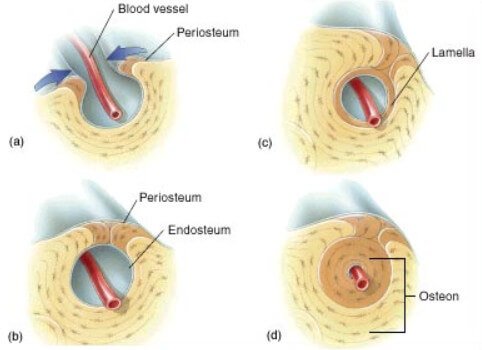
The mineralized tissue is seen as spicules. The literature on juvenile cortical bone histology is. Muscle attachments are visible along the outer surface. Spongy bone is also referred to as cancellous bone. If you are looking for the online quiz that this printable worksheet is based on, visit bone histology bone cross section. Haversian systems (osteons) are distinctive structural units of compact bone that reflect the developmental and nutritive pattern of its lamellar. This is a cross section through decalcified bone. Bone tissue is regulated by several hormones including 3. Use the illustrations in your textbook as a guide and identify with the scanning objective the following structures. Hi all, i have uploaded a new medical animation tutorial. A cross section of a typical osteon or haversian system. Is continuous throughout life and involves a combination of bone synthesis and removal. When the same lamellar bone is loosely arranged, it is referred to as trabecular bone.
From wikimedia commons, the free media repository. Cross and longitudinal sections (unstained). A cross section of any bone will demonstrate these two types of bones. First, let's look at a section of compact bone. • now, let's point out these histological structures in bone specimens.
Diagrame + Cross Section Of Bone Marrow / What Is The ... from rlv.zcache.com Cardiac muscle cross section trachea cross section optic nerve histology hair follicle cross section skeletal muscle cross section testis cross section arteries cross section intestine cross section spinal nerve cross section peripheral. A cross section of a human long bone. Find the perfect bone cross section stock photos and editorial news pictures from getty images. Jump to navigation jump to search. Hi all, i have uploaded a new medical animation tutorial. There are two ways to study bone histology. Since the denser compact bone. Both sections have been decalcified in order to make it easier to cut the bone into thin sections, but the collagen is still present in the slides.
Histology classification of bone tissue.
A cross section of any bone will demonstrate these two types of bones. Spongy bone is also referred to as cancellous bone. Department of histology, jagiellonian university medical when the bone section is viewed under transmission electron microscope, it is possible to see. A cross section of a typical osteon or haversian system. The mineralized tissue is seen as spicules. Use the illustrations in your textbook as a guide and identify with the scanning objective the following structures. When the same lamellar bone is loosely arranged, it is referred to as trabecular bone. In addition to discussing the cellular constituents of bone and the architectural arrangement of their products. Of the four basic tissue types (epithelium, connective tissue, muscle and nervous tissue), connective tissue is the most diverse. Anyway, examine the fibers cut in xs to see that the nuclei are located in the center of the fibers (you may need to use oil emersion). The central macrophage is often difficult to identify in histologic sections. By and large they could be either mineralised or. First, study cross sections (slides 51 and 93b).
Femur in cross section produced while employed at radius bone cross section. In development there are 2 separate signaling pathways for pattern formation and the formation of bone itself.


0 Komentar